Embryology
Bill Heald
Joep Knol
It is fascinating to see how a simple tube-shaped GI-tract evolves into a complex organ system in the adult. However, final embryonic relationships remain simple.

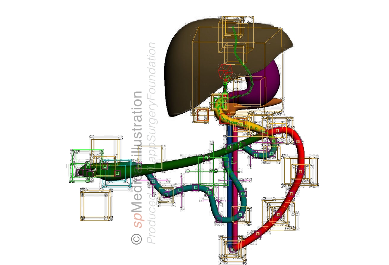
The primitive GI-tract consists of three parts, each with its own main artery:
° Foregut - Celiac trunk
° Midgut - Superior mesenteric artery
° Hindgut - Inferior mesenteric artery
The main reasons for the complexity in adult life are:
1. Rotation of the abdominal foregut tube: 90° clockwise
2. Development of the greater omentum and lesser peritoneal sac from the dorsal mesentery of the abdominal foregut
3. Rotation of the midgut 270° around the superior mesenteric artery
4. Impressive growth of the midgut intestine
Hindgut
The distal colon, rectum and the anal canal above the dentate line are all derived from the hindgut. Therefore this segment is supplied by the inferior mesenteric artery, with corresponding venous and lymphatic drainage.
Its parasympathetic outflow comes from S-2, S-3, and S-4 via splanchnic nerves
Development
5 weeks
Before the fifth week of development, the intestinal and urogenital tracts terminate in a common channel - the cloaca.
5 weeks
Expansion and early rotation of the stomach. Intestinal loops begin to form.
6-8 weeks
There are prominent intestinal loops and rotation of the stomach is completed.
The urorectal septum or fold of Tourneux migrates caudally and divides the cloacal closing plate into an anterior urogenital plate and a posterior anal plate.
8 weeks
Because of rapid growth of midgut, it extends into the umbilical cord and starts rotating around the superior mesenteric artery. Also, the foregut rotates 90° around its axis as the enlarging liver moves to the right
10-12 weeks
Intestines have returned to the abdominal cavity. They push the ascending and descending colon against the body wall.
Embryology and rectal surgery
Due to the processes of embryological development the gut mesentery is one continuous organ from the ligament of Treitz to the rectum. This leads to the opportunity to separate the lateral and retroperitoneal structures from the mesenteric pedicle package in the middle.

Because gastrointestinal cancers tend to progress within this midline pedicle before invading beyond it, purposeful dissection around this pedicle is important for both colon and rectal cancer surgery. The fascia surrounding the mesorectum seems to be an important oncological barrier to the spread of tumor cells.
Rectum
In rectal cancer surgery the whole concept of the mesorectum and dissection according to the avascular plane is based on embryology.
Bill Heald introduced terms like the “holy plane” and “stay on the yellow side of the white” to show how the midline mesorectal envelope can be removed intact in a TME-procedure (Total Mesorectal Excision).
