Anatomy and Embryology
Ayhan Kuzu
Joep Knol
The retroperitoneum forms the posterior plane of dissection in all colectomies and can be divided in different compartments. Understanding of these compartments and intercommunication between them originates from dissection of cadavers.

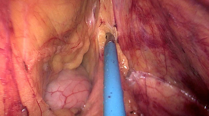
Dr. Ayhan Kuzu demonstrates anatomical structures in a cadaveric dissection
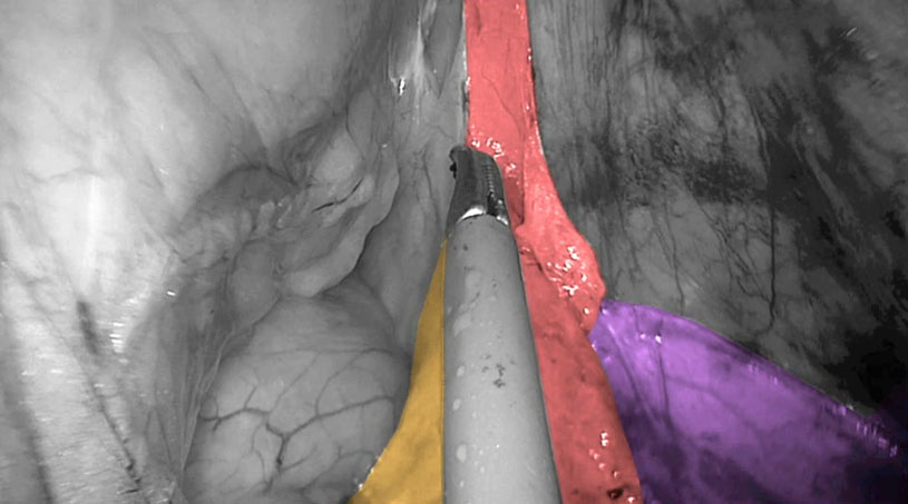
The complexity of the developing mesocolon transversum, interfascial plane and fusion fascia is investigated on human fetuses. Focusing on the splenic flexure it seems that the peritoneum covering the pancreas is much thinner than at other sites and fused with the transverse mesocolon as a “fusion fascia”. This was already described by Carl Toldt in 1879.


Demonstration of fusion fascia of Toldt in red, pancreas in yellow and Gerota’s fascia in purple (Copyright Dr. Joep Knol)
The inferior mesenteric vein provides a peritoneal fold (in fetuses at 20-30 weeks) bridging the peritoneal cavity near the duodenojejunal junction.
Understanding of the different anatomic compartments of the abdominal cavity can guide submesocolic dissection.