Laparoscopic Ultrasound of the Liver
Allessandro Ferrero
Laparoscopic liver surgery is an emergent technique, increasingly performed throughout the world thanks to evolution of technology, improved surgeons experience and the standardization of some procedures. An increasing number of reports shows that laparoscopic hepatectomy has potential advantages over open surgery in terms of blood loss, patient comfort and postoperative hospital stay [1, 2].
IOUS
Intraoperative ultrasonography (IOUS) is a pillar of liver surgery, with an established role to complete disease staging, for resection planning and guidance to liver resection [3, 4].
If laparoscopic liver surgery has the ambition to compete with the open technique, it has to employ IOUS with the same performance.
LUS
Laparoscopic ultrasound (LUS) use was first reported in 1981 by Fukuda et al. [5]. In spite of this early introduction, it has been poorly developed and studied. Nonetheless LUS is reported to increase surgical safety [6,7], and its reliability for staging liver diseases was demonstrated by its performances similar to those of open IOUS [8].
Furthermore, it has to provide real-time guidance during surgery. A peculiar role of ultrasound in laparoscopy is to overcome technical difficulties implicit of the laparoscopic approach, such as lack of palpation, loss of spatial orientation, lack of depth perception that can impair the accuracy of the transection plane.
LUS is often referred to as an important technique for laparoscopic liver resections, but few authors described LUS technique [9], which is far from being standardized.
As in open surgery several subsequent LUS explorations are needed.
Understand anatomy
The first has the purpose to study liver anatomy in a standardized sequence (caval confluence, portal bifurcation, left segmental pedicles, right segmental pedicles, hepatic pedicle), in order to understand the patient specific anatomy and to rule out anatomical anomalies.
Disease stadiation
Then the liver is scanned to study liver parenchyma and its lesions, in order to provide the ultimate and more accurate disease stadiation. When all the lesions (and sometimes new lesions) have been identified, LUS is again performed in order to guide the resection.
LUS is undoubtedly more complex than IOUS, so we schematized our standard technique [10], which consists in creating a map of the liver under ultrasound guidance, in a 5 steps method.
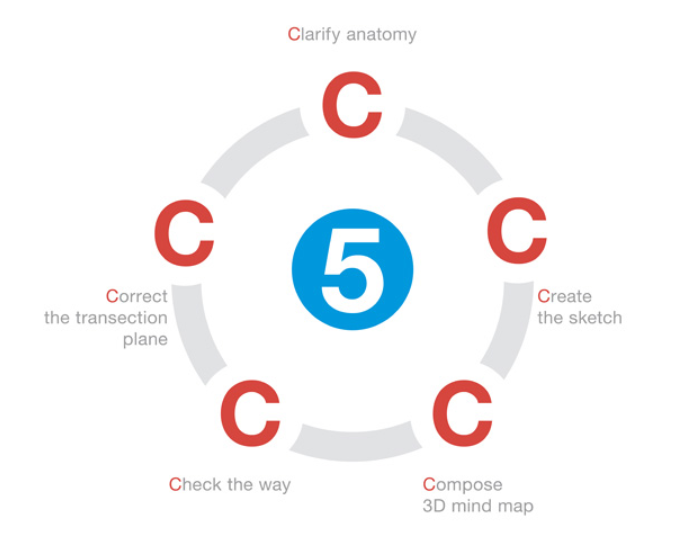
The first step consists in the clarification of anatomy.
The relationships between tumor and surrounding vessels are explored.
Hepatic veins, portal pedicles and their branches, directly involved or just surrounding the lesion are visualized and recognized.
In case of one of the most frequent laparoscopic resections, the left lateral sectionectomy, the surgeon searches for the glissonian pedicles of segment 2 and 3 arising from the umbelical portion as well as the branches of the left hepatic artery.
Another landmark is the middle and left hepatic vein joining in the common trunk. The scissural vein has to be searched as well and its confluence in the middle or left hepatic vein has to be identified.
The second step consists in the creation of a sketch of the underlying anatomical structures on the liver surface with the cautery.
When a vessel can be scanned in the longitudinal plane, a mild compression of the probe will leave a brief impression in the glissonian capsule that can be marked with cautery. When vessel is cross scanned, it can be targeted with the artifact of the hook slipped beneath the US probe and then properly marked. A series of cross-section marks can be joined to draw the long axis of the vessel.
Aim of the sketch is to help the surgeon to create and hold in mind a three-dimensional map of the liver anatomy in relation with the lesion.
The resection will be guided by this mind map and lines of transection are drawn according to the map previously marked, thus establishing which vessel will be ligated and cut and which will be preserved and exposed on the cut surface.
This way the sections lines run exactly where the surgeon will come across vascular structures and divide them.
This is not enough to conduct a safe laparoscopic (and open) liver resection, because the map sketch shows just the glissonian projection of deeper structures.
Therefore the fourth step consists in checking the way of the section plane while on proceeding with the transection.
The resection line is easily visualized as an inhomogeneous hyperechoic linear artifact in the parenchyma, so in any moment the surgeon can check the plane of resection concerning the relationships with the hepatic veins, portal pedicles and surgical margin.
Hepatic veins are usually good landmarks to follow within the liver to ensure the right depth of the transection plane. Repeated assessment of the direction is an easy and safe method to reach the veins to follow and expose, avoiding damages.
During the same operation the surgeon can verify that the pedicles found during the transection are really the ones that need to be divided. During the resection is of course mandatory to check the correct surgical margin in order to perform a safe oncological operation.
The resection plane is constantly verified by ultrasound, in order to maintain a safe margin, providing surgeon with immediate feedback of possible changes.
The direction is in fact not always right from the beginning, so the fifth step consists in correcting the direction of the section line whenever needed.
It is not so obvious to start the resection with the right angle of incidence and often the direction has to be adjusted in order to stay clear of the lesion, to reach a pedicle at the correct distance from its origin or to reach a structure to spare and follow.
Ultrasound offers the chance to correct the plane of transection when on the wrong way, in order to perform liver resection safely.
Conclusion
In conclusion, laparoscopic liver ultrasound meets the surgeon needs, concerning staging, planning and above all guidance to liver transection.
The technique we developed, which we called US liver map technique, can increase the safety of laparoscopic hepatectomies, showing always the way to go during the transection.